Memory loss, tremors, paralysis: when parts of the nervous system start to break down – or get broken – the consequences for human health can be staggering. Can we fix the nervous system, and how are scientists approaching the problem? We take a deep dive into various strategies for interfacing with the nervous system to restore neuronal function.

If you’ve accidently cut yourself – a minor cut – then your body would likely heal itself by generating new skin cells at the wound in a phase of healing called proliferation. It’s a whole other story if you cut off a body part. Unlike salamanders who can grow back their tails, we humans are unable to regenerate body parts, even relatively small parts like a finger. That’s because the cells responsible for generating fingers, so-called stem cells, are only actively growing whole fingers during embryotic development.
Similarly, our bodies have a limited ability to heal damage to the nervous system because the stem cells responsible for growing a functioning nervous system are likewise only fully active in the embryo. If you were to zoom into parts of the nervous system, you would see a network of billions of interconnected cells called neurons, the fundamental building blocks of the nervous system responsible for transmitting electrical signals throughout the body. The number of neurons in the body peaks before birth, at roughly 86 billion units, and slowly declines throughout one’s lifetime.
That doesn’t mean that new neurons can’t be made. There is evidence that points to neuron birth in specific regions of the brain, albeit at a slower rate as we age. But unlike skin cells that can regenerate to heal a small wound, there is no way of spontaneously growing new neurons to heal a lesion of the nervous system. So how do you fix a damaged nervous system if the body can’t heal the lesion with new neuron cells?
The importance of neuroplasticity
“In the brain, there is no regeneration or repair, but neuroplasticity,” says Defitech Chair of Clinical Neuroengineering Friedhelm Hummel, who specializes in noninvasive deep brain stimulation. “Rehabilitation is about getting neurons to rewire their branches and make connections across a lesion. The nervous system’s capacity to adapt is called neuroplasticity.”
Neuroplasticity is what gives us the ability to learn new information and adjust to new situations. From childhood to adulthood, those 86 billion neurons that we are born with are constantly firing electrical signals, connecting and rewiring as we learn and adapt, and they do so thanks to the various branches that extend from the neuron’s cell body. The branches that transmit signals from one neuron to the next are called the axons, and those that receive signals from another neuron are called dendrites. In other words, neuroplasticity is the ability of these branches to change the way they connect to each other, essentially adjusting the way the network of neurons fire and transmit electrical signals.

When neurons are no longer functional, or die, neuroplasticity will attempt to rewire the surrounding, intact neuron branches to reestablish communication channels. For large lesions, like blunt trauma or illness or a spinal cord injury leading to paralysis, the gap may simply be too big for neuroplasticity alone to renew meaningful communication channels.
At EPFL, researchers, engineers, doctors and scientists are exploring ways to restore communication pathways of the damaged nervous system, be it in the brain, the spinal cord or the peripheral nervous system. The nervous system may still output useful information, and may also be capable of receiving input thanks to artificial stimulation of the nervous system, both fundamental in the development of rehabilitation protocols of the nervous system for translation into meaningful clinical therapies with life-changing potential.
Prosthetic approach to rehabilitation
Current state-of-the-art prosthetic technology for neurorehabilitation involves interfacing the nervous system with surgically implantable electrodes, usually printed on a flexible material, directly in contact with the brain or the rest of the nervous system. The noninvasive approach to rehabilitation involves placing an electronic device, such as an electrode, on the skin to deliver signals to the nervous system. The pharmaceutical approach involves the use of drug therapy to increase neuroplasticity conducive to learning new tasks. Then there is iontronics, a system based on ion transport instead of electrons, developed at EPFL by Yujia Zhang. As an emerging approach for neurorehabilitation, researchers are exploring ways to communicate with the nervous system by controlling the movement of ions and small molecules.
Neuroprosthetics are devices that aim to restore lost or impaired neural function by interacting with the nervous system. The role of neuroprosthetics is to replace sensory or motor functions, or modulate brain activity.
“There is no one-size-fits-all solution,” explains Silvestro Micera, head of EPFL’s Translational Neural Engineering Laboratory and neuroengineer at both EPFL and Scuola Sant’Anna. His specialty is in the restoration of hand sensory-motor control in people with different disabilities. “Where you interface with the nervous system depends on the function you want to restore, the neurophysiology of that function, and the specifics of the patient’s lesion.”
Among Micera’s expertise is the development of neuroprosthetics that restore touch sensation in hand amputees by interfacing with the peripheral nervous system, specifically the nerves in the arm. “We can simulate the sensation of touch from the missing hand by electrically stimulating the residual nerves in the arm. In practice, we’ve been interfacing with rather large nerves, so we’ve opted to use intraneural electrodes to deliver the stimulation in order to intercept multiple nerve bundles to simulate sensory feedback from the missing hand.”
“Where you interface with the nervous system depends on the function you want to restore, the neurophysiology of that function, and the specifics of the patient’s lesion.”
– Silvestro Micera, professor at EPFL’s Translational Neural Engineering Laboratory
Intraneural electrodes are essentially minute electrode arrays, less than 0.3 mm by 3 mm in size, that traverse a section of the nerve fiber. The insertion of intraneural electrodes requires precision neurosurgery and has been successfully performed on amputees in collaboration with Italian partners. Lately, Micera and colleagues have been working to repurpose the intraneural electrodes to make them capable of delivering electrical impulses to restore hand function in people with spinal cord injury.
Neuroscientist Grégoire Courtine and neurosurgeon Jocelyne Bloch, both at EPFL and the Lausanne University Hospital (CHUV), and co-founders of NeuroRestore, are developing a “digital bridge,” a neuroprosthetic system that bridges the gap created by lesions disrupting signals between the brain and the rest of the body, such as in cases of paralysis.
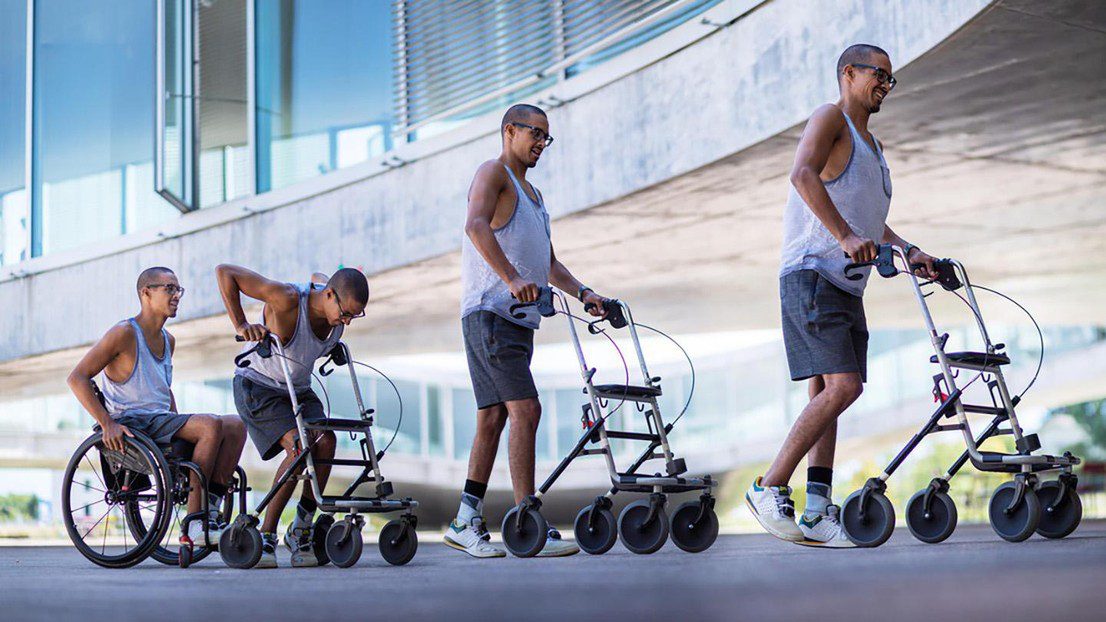
“With our digital bridge, we are translating the paralyzed patient’s intention to move into action,” explains Courtine. “We have successfully helped five individuals who were paralyzed after an accident: three who were able to walk again, and two who were able to move their arms,” says Bloch.
Their digital bridge strategy involves interfacing the patient’s brain to detect brain signals, and translate them to the affected parts of the body such as the arms or the legs via the spinal cord.
At the interface of the brain, Courtine and Bloch are using electrodes, about 5 cm in diameter, which are surgically implanted at the surface of the brain. “I like to call these electronic bones. We simply remove part of the skull, just above the brain region that controls the legs, and replace it with the electronic bone that will listen to those brain cells,” explains Bloch.
Electrical stimulation of the spinal cord
At the spinal cord interface, the duo has opted for a flexible electrode array, about 1 cm by 6 cm in size, developed by Courtine and Bloch’s spin-off ONWARD Medical. This array is expertly inserted by Bloch beneath the vertebrae and wraps around the back of the spinal cord.
ONWARD Medical has recently obtained FDA approval to commercialize their spinal cord stimulation technology in the United States. “It’s the first time in the history of humanity that a therapy has been approved to improve rehabilitation after a spinal cord lesion,” says Courtine.
Electrical stimulation of the spinal cord has also proven useful for treating patients suffering from Parkinson’s disease. “We looked at a group of patients who had tremendous difficulty walking. We applied the same principle of spinal cord stimulation, this time without the digital bridge, and we were able to correct deficits in the patients’ gait and reduce the rate of falling,” says Bloch.
Courtine and Bloch have also investigated use of deep brain stimulation probes, specifically of the lateral hypothalamus, and found improved recovery of lower limb movements in two individuals with partial spinal cord injury.
Most electrodes that interface with the human body – such as the ones used by Micera, or Courtine and Bloch – consist of a circuit printed on a flexible polymer, which despite being flexible, is still relatively rigid compared to the organic nature of the nervous system. Stéphanie P. Lacour, an interdisciplinary neuroengineer at EPFL, is developing a whole new field of stretchable electrodes. She made a breakthrough discovery about stretchable metal films and their applications in soft devices. “I was exploring how to design electrodes that could conform to objects of irregular curvature. The first idea was to deposit metal on a compliant polymer carrier. I started with gold, a ductile metal, and silicone, an elastomer. To my surprise, the metal could be evaporated on the silicone, was electrically conductive, and could retain its conductivity when stretched.”
“With our digital bridge, we are translating the paralyzed patient’s intention to move into action.”
– Grégoire Courtine, professor and director of the EPFL Neuro-X Institute and co-founder of NeuroRestore
Driven to connect these stretchable electrodes with biology, she has since been developing innovative, stretchable electrodes at the intersection of robotics: with deployable electrodes that open like a flower 4 cm across to ensure maximum coverage on the surface of the brain while passing through a minimally invasive 1 cm hole in the skull; to auditory implants that closely conform to the curved surface of the brainstem for high-resolution prosthetic hearing; to enhancing electrodes that could interface with potentially any part of the nervous system.
Noninvasive approach to rehabilitation
For treating brain injury, Hummel is investigating ways to stimulate deep structures within the brain and he decided years ago to explore noninvasive solutions. “Deep brain stimulation with a probe is the most established interface for the brain, and yet only two to four percent of Parkinson’s patients can benefit from it,” he explains. “In contrast, noninvasive brain stimulation has the potential to reach a large number of patients.”
By tuning electrical signals delivered via electrodes placed on a patient’s head, Hummel is able to target deep structures within the brain. “Neurons respond to low frequency signals, between one and one hundred Hertz, yet remain unresponsive to high frequency signals in the kilo Hertz range. We’ve taken advantage of these characteristics to target and stimulate very precise regions of the brain, located with the help of magnetic resonance imaging and computational modelling,” explains Hummel.
“In humans, we’ve demonstrated that our non-invasive deep brain stimulation enhances plasticity of the targeted deep brain area,” explains Hummel. Although rehabilitaion studies have yet to be published, several clinical trials are ongoing to demonstrate the potential for improving motor and cognitive functions in impaired populations, such as stroke and traumatic brain injury.”
Micera and his research team have also been exploring non-invasive technology for restoring thermal sensation in amputees. By delivering hot and cold directly on the amputee’s residual arm through a specialized interface, the researchers were able to restore sensations of warmth and cold in the missing hand.
Pharmaceuticals and neuroplasticity
Drug therapy may capitalize on the nervous system’s incredible ability to adapt, specifically its neuroplasticity. Courtine, Bloch and team are exploring how gene therapies may promote nerve growth after spinal cord injury in animal models. The scientists activated growth programs in the identified neurons in mice to regenerate their nerve fibers, upregulated specific proteins to support the neurons’ growth through the lesion core, and administered guidance molecules to attract the regenerating nerve fibers to their natural targets below the injury. Mice with anatomically complete spinal cord injuries regained the ability to walk, exhibiting gait patterns that resembled those quantified in mice that resumed walking naturally after partial injuries.

Iontronics, communicating with the language of cells
For the past two decades or so, researchers around the world have been exploring how to communicate with the nervous system, and printed circuits of electrodes have been used time and again in neuroprosthetics. But electrodes use electrons to produce signals, while neurons use a complex biological mechanism based on ion movements. For example, essential ions used in cell function include potassium and sodium, which are positively charged ions actively controlled by cell membranes to form the molecular basis for all cellular activities. Yujia Zhang, who leads EPFL’s Laboratory for Bio-Iontronics, is pioneering the development of droplet-based ionic devices in the emergent field of iontronics.
Electrodes are electronic conductors, and reactions at the electrode-tissue interface are required to mediate the transition from electron flow in the electrode-to-ion flow in the tissue. “Electrodes are inefficient at interfacing with the nervous system. High currents are used to counteract the effect of ion accumulation on the electrodes, known as the electric double layer, which can decrease stimulation efficacy. So, I’ve been exploring ways to develop biocompatible bio-inspired electronics to overcome this issue,” explains Zhang. He and his team are deploying microfluidic technology to print miniature biocompatible droplet-based iontronic devices, termed dropletronics, which include iontronic diodes, transistors and logic gates, the building block analogues of electronic components. One iontronic transistor measures roughly 250 micrometers in size. “Our iontronic transistor can serve as a biocompatible sensor to record ion movements from sheets of human cardiomyocytes, revealing their beating patterns. Our dropletronics will pave a way to the assembly of miniature bioiontronic systems,” explains Zhang.
This article was published in the June 2025 issue of Dimensions, an EPFL magazine that showcases cutting-edge research through a series of in-depth articles, interviews, portraits and news highlights. Published four times a year in both English and French, it can be sent to anyone who wants to subscribe as well as contributing members of the EPFL Alumni Club. It is also distributed free of charge on EPFL’s campuses.
Author: Hillary Sanctuary
Source: EPFL